Jump to top menu Jump to main menu Jump to content
Core Facility

AMIE - Applied Molecular Imaging Erasmus MC

AMIE is a Core Facility Research with state-of-the-art imaging technology. We are an accessible infrastructure, training center and expert center regarding preclinical and translational imaging techniques with dedicated staff and management.

Services

Methods / applications

Erasmus MC has been investing in imaging expertise and systems for quite some time and has built unique expertise in a number of areas. Various technologies (i.e. equipment as well as expertise) are put together in one new facility: Applied Molecular Imaging Erasmus MC. Combinations of imaging modalities can be applied to a single research question, or one experimental animal. From experience we know that a physical grouping of complementary techniques and expertise is a must to reach successful synergy. The new central imaging facility is largely housed in the new research unit in the Erasmus MC Experimental Animal Centre (EDC).

The research in the departments of Erasmus MC covers a wide range of topics; cancer research, microbiology/immunology, developmental biology and pharmacology.

Equipment

Discover our comprehensive selection of state-of-the-art preclinical imaging equipment designed to enhance your research capabilities:

  • Discovery MR901 – 7T MRI
  • FMT2500 – Fluorescent Optical Imaging
  • Gammacounter for radioactive samples
  • IVIS Spectrum – Optical Imaging
  • MSOT – 3D Optoacoustics
  • NanoScan – SPECT, 1T MRI
  • Quantum Gx2 – microCT
  • VECTor – SPECT/PET, microCT and Optical Imaging
  • Vevo3100 - Ultrasound
  • Vevo LAZR-X photoacoustic addition to the Vevo3100
  • Workstation with analysis software packages Imalytics, PMOD, Analyze, VEVOlab and more 

Apparatuur_AMIE

Showcases

Explore our showcases of cutting-edge preclinical imaging, where groundbreaking techniques and applications come to life, advancing our understanding of complex biological processes and disease mechanisms.

1. Quintuple Modality Tumor Necrosis Imaging

"Unlocking Insights into Tumor Aggressiveness and Therapy Effectiveness"

Leverage the power of both nuclear and optical imaging to pinpoint therapy-induced tumor necrosis. This multimodal approach offers complementary insights into tumor response. PET with FDG closely monitors tumor metabolism and serves as a benchmark for treatment efficacy. While SPECT and fluorescent imaging (FLI), armed with an advanced necrosis-avid contrast agent, accurately reveal areas of necrosis, exploiting damaged membrane integrity to image cell death. Additionally, bioluminescent imaging (BLI) detects viable tumor cells. Together, this comprehensive array of imaging modalities provide vital insights guiding tailored treatment strategies. 

This figure was originally published in: 
Stroet MCM, de Blois E, de Jong M, Seimbille Y, Mezzanotte L, Löwik CWGM, Panth KM. Improved Multimodal Tumor Necrosis Imaging with IRDye800CW-DOTA Conjugated to an Albumin-Binding Domain. Cancers. 2022; 14(4):861.

2. Optical Imaging of Tumor Response

“Explore the Impact of Therapy on Tumor Vasculature”

Optical imaging provides a non-invasive way to monitor processes involved in tumor growth and metastasis formation in immunodeficient mice. Here fluorescence molecular tomography (FMT), with its advanced 3D imaging capabilities, is used to delve into the effects of hyperbaric oxygen therapy (HBOT) and irradiation on tumor dynamics. By analyzing blood vessel quality with AngioSense and unraveling hypoxia with HypoxiSense probes, FMT offers a comprehensive overview of the tumor environment. Seamlessly integrating FMT with CT images provides precise anatomical references.  Thus, optical imaging methods can unveil longitudinal insights into tumor characteristics, revolutionizing oncology research.
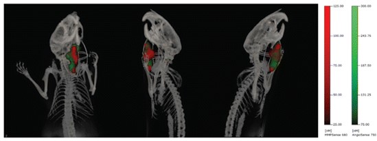
This figure was originally published in: 
Braks JAM, Spiegelberg L, Koljenovic S et al. Optical Imaging of Tumor Response to Hyperbaric Oxygen Treatment and Irradiation in an Orthotopic Mouse Model of Head and Neck Squamous Cell Carcinoma. Mol Imaging Biol 17, 633–642 (2015).
 

3. Imaging Drug Delivery with SPECT and DCE-MRI

“Unveiling Tumor Dynamics”

The combination of SPECT and MRI can unveil vital insights into the perfusion efficiency and the accumulation of radiolabeled receptor-targeting peptides in solid tumors. This multimodal approach reveals nuanced variations in functional tumor characteristics, even in seemingly homogeneous tissue. By leveraging DCE-MRI-derived parameters, it is possible to decode tumor perfusion, vessel density, and permeability, unlocking insights into the impact of vascular traits on the efficacy of receptor-targeting therapies. 

This figure was originally published in: 
Haeck JC, Bol K, de Ridder CMA et al. Imaging heterogeneity of peptide delivery and binding in solid tumors using SPECT imaging and MRI. EJNMMI Research. 2016; 6(3).

Access & costs

Access to and use of the AMIE instrumentation is permitted under the supervision of AMIE personnel. Training is provided for those who plan to use the equipment independently.

Costs for equipment use depends on user requirements. For more information contact the AMIE.

Support

The AMIE provides full-service support on all equipment for imaging experiments, with the option to obtain training for independent use.

All imaging modalities have isoflurane equipment for anesthesia.

Training & education

Training

Those interested in conducting their own imaging research can undergo training to operate the equipment, develop optimized imaging protocols tailored to their research question, and receive instructions on data analysis techniques.

Education

AMIE is involved in several educational activities including workshops (e.g., Translational Imaging Workshop by AMIE ‘From mouse to man’), symposia (e.g., OIC & AMIE Advanced imaging Techniques; From bench to bedside) and Masters programs (e.g., Infection & Immunity, Neuroimaging).

As the imaging field continues to expand, we believe in the importance of ongoing development and learning. Therefore, we are currently exploring options to organize in-depth workshops focused on specific imaging techniques, aimed at enhancing both skills and knowledge of our users and anyone interested.

If you are seeking a lecture or talk on preclinical imaging techniques and/or applications for your program, or if you would like to arrange a guided tour through the AMIE facility, please do not hesitate to reach out to us via email.

News, events & publications

News

Register now for our upcoming Translational Imaging Workshop by AMIE ‘From mouse to man’ - April 7th - 9th 2025

Register now using the QR code on the flyer or by clicking this link


Impression of our 'Data Analysis Techniques for Preclinical Imaging' workshop - November 28th & 29th 2024

Last November, the AMIE core facility together with the Imaging office of the Erasmus MC, organized a workshop focused on Data Analysis Techniques for Preclinical Imaging. We provided the participants with essential insights and skills for analyzing preclinical imaging data and the opportunity to network with fellow researchers and industry.

We were able to do this through the enthusiastic involvement of experts like Chris Vanhove (Image processing basics), Ilva van Houwelingen (Advanced image processing techniques + hands-on training with freeware: Weasis and 3D slicer), Theo van Walsum (hands-on training with freeware: MeVisLab), Oleh Dzyubachyk (Analysis of small animal imaging data: examples, challenges, and opportunities) and Joanes Grandjean (Data curation). Felix Gremse from Gremse-IT and Geoffrey Warnock from PMOD Technologies provided a hands-on training with commercial available data analysis software (Imalytics and PMOD, respectively). 

This photo collage gives you an impression of the workshop:


Launch of AMICE national platform

AMIE is a founding member of AMICE (Advanced Multi-modal Imaging CEnters). AMICE is a Dutch national platform in which preclinical imaging centers participate for full-scale, collective and synergistic collaboration within and between centers. AMICE harbors a wide range of state-of-the-art preclinical imaging equipment and expertise.

AMICE aims to reinforce the preclinical imaging infrastructure by coordinating access to imaging equipment and by building a shared network of expertise, people, equipment and data.


Impression of our Translational Imaging Workshop by AMIE ‘From mouse to man’ - November 20th - 22nd 2023

We organized a three day Translational Imaging Workshop “From Mouse to Man” and had put together a program that gave a broad introduction to the exciting field of preclinical and molecular translational imaging and their applications in biology and medicine. The workshop covered a wide array of imaging technologies, including:

  • Nuclear imaging (PET and SPECT)
  • Magnetic resonance imaging (MRI)
  • Computed tomography ((µ)CT)
  • Fluorescent imaging (FLI)
  • Bioluminescent imaging (BLI)
  • Ultrasound imaging (US)
  • Photoacoustic Imaging (PAI)

Next to the efforts of the AMIE team, we were able to do this through the enthusiastic involvement of expert researchers. We are very grateful for the involvement of Sanne van Lith, Louise Van der Weerd, Uwe Himmelreich, Frans Meijers, Freek Beekman, Christian Vanhove, Laura Mezzanotte, Clemens Lowik, Hans Bosch, Klazina Kooiman, Anton Nikolaev, Francis Kalloor Joseph, Marieke Stammes and Amanda Kiliaan. In addition to the opportunity to learn about the many different modalities and their applications, our workshop also offered an excellent possibility to network with other researchers and representatives from industry. We could offer these networking events, as lunches and a dinner on Monday, thanks to the sponsorship of BIOEMTECH, MILabs, UNO Life Science Solutions, Revvity, MOLECUBES NV, FUJIFILM VisualSonics, Inc., PD Dr. Felix Gremse, iThera Medical GmbH and MR SOLUTIONS GROUP.

See the photo collage below for an impression of our venue and workshop:


Events

Conferences

Stay informed and inspired at these upcoming imaging conferences:

  • European Symposium on Ultrasound Contrast Imaging, Rotterdam, The Netherlands, January 16-17, 2025
  • NKRV Workshop, Nederlandse Klinische Radiochemische Vereniging, Rotterdam, The Netherlands, January 31, 2025
  • EMIM, European Molecular Imaging Meeting, Bilbao, Spain, March 11-14, 2025
  • SNMMI annual meeting, Society of Nuclear Medicine and Molecular Imaging, New Orleans, Louisiana, USA, June 21-24, 2025
  • WMIC, World Molecular Imaging Congress, Anchorage, Alaska, September 29 - October 3, 2025
  • EANM annual meeting, European Association of Nuclear Medicine, Barcelona, Spain, October 4-8, 2025
  • ESMRMB annual scientific meeting, European Society for Magnetic Resonance in Medicine and Biology, Marseille, France, October 8-11, 2025

Publications and theses

Recent publications

Explore the cutting-edge research enabled by our state-of-the-art imaging equipment in the latest publications from our esteemed users:


The Balance Between the Therapeutic Efficacy and Safety of [177Lu]Lu-NeoB in a Preclinical Prostate Cancer Model
Verhoeven M, Haeck J, de Blois E, Orlandi F, Barbato D, Tedesco M, Konijnenberg M, Dalm SU. 2024. Molecular Imaging and Biology, Feb;26(1):114-123.
https://doi.org/10.1007/s11307-023-01851-4


The association between human blood clot analogue computed tomography imaging, composition, contraction, and mechanical characteristics
Cruts JMH, Giezen JA, van Gaalen K, Beurskens R, Ridwan Y, Dijkshoorn ML, van Beusekom HMM, Boodt N, van der Lugt A, de Vries JJ, de Maat MPM, Gijsen FJH, Cahalane RME. 2023. PLoS One, Nov 13;18(11):e0293456.
https://doi.org/10.1371/journal.pone.0293456


Side-by-side comparison of the two widely studied GRPR radiotracers, radiolabeled NeoB and RM2, in a preclinical setting
Damiana TST, Paraïso P, de Ridder C, Stuurman D, Seimbille Y, Dalm SU. 2023. European Journal of Nuclear Medicine and Molecular Imaging, Nov;50(13):3851-3861.
https://doi.org/10.1007/s00259-023-06364-4


Bioluminescence imaging on-chip platforms for non-invasive high-content bioimaging
Araújo-Gomes N, Zambito G, Johnbosco C, Calejo I, Leijten J, Löwik C, Karperien M, Mezzanotte L, Teixeira LM. 2023. Biosens Bioelectron, Oct 1;237:115510.
https://doi.org/10.1016/j.bios.2023.115510


Synthesis and Evaluation of ePSMA-DM1: A New Theranostic Small-Molecule Drug Conjugate (T-SMDC) for Prostate Cancer
Murce E, Spaan E, Beekman S, van den Brink L, Handula M, Stuurman D, de Ridder C, Dalm SU, Seimbille Y. 2023. Pharmaceuticals (Basel), Jul 28;16(8):1072.
https://doi.org/10.3390/ph16081072


Addition of heparin binding sites strongly increases the bone forming capabilities of BMP9 in vivo
Siverino C, Fahmy-Garcia S, Niklaus V, Kops N, Dolcini L, Misciagna MM, Ridwan Y, Farrell E, van Osch GJVM, Nickel J. 2023. Bioactive Materials, Jul 18;29:241-250.
https://doi.org/10.1016/j.bioactmat.2023.07.010


Whole-body bioluminescence imaging of T-cell response in PDAC models.
McMorrow R, Zambito G, Nigg A, Lila K, van den Bosch TPP, Lowik CWGM, Mezzanotte L. 2023. Frontiers in Immunology, Jul 11;14:1207533.
https://doi.org/10.3389/fimmu.2023.1207533


[212Pb]Pb-eSOMA-01: A Promising Radioligand for Targeted Alpha Therapy of Neuroendocrine Tumors
Chapeau D, Koustoulidou S, Handula M, Beekman S, de Ridder C, Stuurman D, de Blois E, Buchatskaya Y, van der Schilden K, de Jong M, Konijnenberg MW, Seimbille Y. 2023. Pharmaceuticals (Basel), Jul 10;16(7):985.
https://doi.org/10.3390/ph16070985


Community Survey Results Show that Standardisation of Preclinical Imaging Techniques Remains a Challenge
Tavares AAS, Mezzanotte L, McDougald W, Bernsen MR, Vanhove C, Aswendt M, Ielacqua GD, Gremse F, Moran CM, Warnock G, Kuntner C, Huisman MC. 2023. Molecular Imaging and Biology, Jun;25(3):560-568.
https://doi.org/10.1007/s11307-022-01790-6


Radiochemical and Biological Evaluation of 3p-C-NETA-ePSMA-16, a Promising PSMA-Targeting Agent for Radiotheranostics
Murce E, Ahenkorah S, Beekman S, Handula M, Stuurman D, de Ridder C, Cleeren F, Seimbille Y. 2023. Pharmaceuticals (Basel), Jun 15;16(6):882.
https://doi.org/10.3390/ph16060882


Preclinical Evaluation of a PSMA-Targeting Homodimer with an Optimized Linker for Imaging of Prostate Cancer
Murce E, Beekman S, Spaan E, Handula M, Stuurman D, de Ridder C, Seimbille Y. 2023. Molecules, May 11;28(10):4022.
https://doi.org/10.3390/molecules28104022


Pre- and Intraoperative Visualization of GRPR-Expressing Solid Tumors: Preclinical Profiling of Novel Dual-Modality Probes for Nuclear and Fluorescence Imaging
Verhoeven M, Handula M, van den Brink L, de Ridder CMA, Stuurman DC, Seimbille Y, Dalm SU. 2023. Cancers (Basel), Apr 5;15(7):2161.
https://doi.org/10.3390/cancers15072161


DNA repair in cardiomyocytes is critical for maintaining cardiac function in mice
de Boer, M.te Lintel Hekkert, M.Chang, J.van Thiel, B. S.Martens, L.Bos, M. M.de Kleijnen, M. G. J.Ridwan, Y.Octavia, Y.van Deel, E. D.Blonden, L. A.Brandt, R. M. C.Barnhoorn, S.Bautista-Niño, P. K.Krabbendam-Peters, I.Wolswinkel, R.Arshi, B.Ghanbari, M.Kupatt, C. … Duncker, D. J. 2023Aging CellMar;22(3):e13768.
https://doi.org/10.1111/acel.13768


Editorial: Small animal imaging: Technological and methodological advances to improve the translational power
Bernsen MR, McDougald W, Mezzanotte L, Moran CM, Tavares A, van der Weerd L. 2022. Frontiers in Medicine (Lausanne), Dec 12;9:1099233.
https://doi.org/10.3389/fmed.2022.1099233

About us

Introduction

Imaging at the AMIE

The AMIE is a facility that houses preclinical imaging equipment, providing researchers with access to specialized tools and the expertise of imaging specialists to address their research questions.


For inquiries please send an email to amie@erasmusmc.nl

Our team

Dr. Monique Bernsen has a PhD in Medical Biology and has been active in Molecular Imaging since 2004. In 2010 she was involved as co-chair in the realization of the AMIE facility, and since 2019 she is Head Operations and Daily Management of the AMIE Core Facility. You can always contact her for questions about the possibilities that AMIE offers for your research and for entering into collaborations. Our goal is to promote and enable the use of advanced preclinical imaging techniques in any way possible.

Dr. Jeroen Essers holds a PhD in Molecular Biology and is a group leader Molecular Imaging at the Erasmus MC. As Head Innovation of the AMIE Core Facility he wants to strengthen the further development of medical and advanced imaging technology and he therefore focusses on exploring, recommending and designing new imaging modalities and programs in order to maintain the pioneering role of the AMIE imaging facility. This way we established a knowledge centre for imaging technology and an accessible test location for new imaging technology. Start-ups can develop innovative imaging technology and present themselves more easily to potential users and investors. Innovation and collaboration are achieved through ease of accessibility of AMIE for internal and external partners.

Dr. Sandra van Tiel completed a BSc in Medical Biology (1995) at the Hoger Laboratorium Onderwijs in Delft and a PhD in cellular imaging (2020) at the Erasmus MC in Rotterdam. Sandra was appointed to her current position in April 2019. Her work primarily involves performing and supervising imaging experiments using PET, SPECT, CT and MRI. Sandra is the registered coordinating radiation expert and the animal coordinator of the AMIE. You can contact her if you need help setting up your experiment, but also if you need help writing a study protocol or project license.

Yanto Ridwan completed a BSc in Medical Biology (2007) at the Hoger Laboratorium Onderwijs in Rotterdam. He was appointed as an application specialist, with the main focus on optical imaging and microCT. His tasks include providing preliminary advice on the use of preclinical imaging modalities, providing technical support and he is also involved in application development, education, planning and equipment maintenance.

Dr. Marjolein Verhoeven completed a MSc in Medical Natural Sciences with a specialization in medical physics (2018) and a MSc in Oncology (2018) at the Vrije Universiteit in Amsterdam. In 2024 she obtained her PhD in preclinical molecular imaging and therapy at the department of Radiology & Nuclear Medicine at the Erasmus MC in Rotterdam. Since 2023, she has been part of the AMIE Core Facility team as an application specialist with a focus on MRI. She is responsible for supporting MRI users through training, instruction and technical assistance, and for expanding the application possibilities of the MRI equipment.

Our expertise

AMIE is uniquely positioned within the Netherlands and relative to facilities in other countries worldwide. Not only does AMIE house an extensive array of imaging equipment with all modalities currently used for preclinical in vivo imaging, AMIE also has expert staff dedicated to the facility and is therefore able to continuously offer research support. Through close interaction with scientific experts and industry, AMIE fulfils a central role in the cycle of advancing imaging techniques, innovative research, and technology development, including hardware, software and wetware. That all equipment is only meters apart or even combined in a single platform is an immense benefit: study objects can be easily transferred between platforms giving investigators the opportunity to use several imaging modalities to gain extensive insight into their research question. This has already resulted in unique discoveries in various fields, e.g. stem-cell, cardiovascular and cancer research, which would not have been possible using single imaging modalities, and there remain many more applications which have yet to benefit from multi-modal experiments. Furthermore, imaging maximizes the information that can be gained from a given animal (refining), contributing to reduction of laboratory animal use.

The unique combination of extensive preclinical imaging equipment, cutting-edge technology, and dedicated in-house experts makes AMIE attractive to researchers both in NL and worldwide.

Collaboration

Discover the diverse range of users and collaborators who have utilized our state-of-the-art facility to push the boundaries of scientific research and innovation:

Erasmus MC departments using the facility:

  • Biomedical Engineering
  • Experimental Cardiology
  • Ear, nose and Throat
  • Hematology
  • Immunology
  • Internal Medicine
  • Medical Oncology
  • Molecular Genetics
  • Neurology
  • Neurosciences
  • Orthopedics
  • Pediatrics
  • Pharmacology
  • Pulmonary Medicine
  • Radiology & Nuclear Medicine
  • Rheumatology
  • (Experimental) Surgery
  • Urology
  • Viroscience

Dutch academic users and partners:

  • Amsterdam Universitair Medisch Centrum (Amsterdam UMC), locatie AMC
  • Biomedical Primate Research Centre (BPRC)
  • Leids Universitair Medisch Centrum (LUMC)
  • Radboud Universitair Medisch Centrum (Radboudumc)
  • Technische Universiteit Delft (TU Delft)
  • Technische Universiteit Eindhoven (TU/e)
  • Universitair Medisch Centrum Groningen (UMCG)
  • Universitair Medisch Centrum Utrecht (UMC Utrecht)
  • Universiteit Maastricht (UM)
  • Wageningen University & Research (WUR)

Dutch non-academic users and partners:

  • Cenya Imaging BV
  • HollandPTC
  • MILabs BV
  • Quirem Medical BV

International academic users and partners:

  • KU Leuven (Prof. Uwe Himmelreich)
  • NCSR Demokritos Athens (Dr. Theodosia Maina-Nock)
  • UGent (Prof. Christian Vanhove, Prof. Julie de Backer)
  • University of Edinburgh (Dr. Adriana Tavares)
  • University of Pittsburgh (Dr. M. Weinberg, Dr. Zsolt Urban)

International non-academic users and partners:

  • Chiesi Pharmaceuticals BV
  • Gremse-IT

Career opportunities

There are currently no available positions.